By Carol Morton
16 February 2021. Location, location, location. That ubiquitous real estate catchphrase now also applies to the latest specialized genomic technologies that may become an important tool in precision medicine.
 Spatially resolved transcriptomics can detect which genes are active in a tissue and in which cells. One method originated in a Swedish laboratory. Last month, the editors of the journal Nature Methods unanimously voted it the Method of the Year 2020. A collection of articles in the January 2021 issue explores the technologies, applications, and future challenges of spatially resolved transcriptomics.
Previously, researchers were able to measure gene expression in single cells, but the cells essentially had to be mixed in a blender, losing essential location information. Now, spatially resolved transcriptomics offers increasing capabilities to evaluate the cells in the context of the tissue in which they function.

One of the two main approaches originated in the Science for Life Laboratory of Joakim Lundeberg at Royal Institute of Technology (KTH) and Jonas Frisén at the Karolinska Institute. A Nature Methods news feature details how the collaboration, led by graduate students Patrik Ståhl and Fredrik Salmén, began in 2009 as a way to extract more information from tissue slides. The team called the resulting technology Spatial Transcriptomics and spun out a company, which was purchased in 2018 by 10x Genomics.
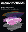
The first paper was published in Science in 2016. The January 2021 cover of Nature Methods depicts the technology.
“Tissues have traditionally been investigated by microscopy capturing the histology, i.e., the morphology, of the samples,” Lundeberg said in an email. “And this has shaped our view of organs and corresponding diseases. With our approach, we can provide a gene activity atlas of the same tissue. This representation is fully unbiased and provides a much higher detail than the microscope image.”
Lundeberg and his colleagues continue to improve the technology and work toward clinical applications in multiple international research collaborations.
They are creating atlases of organs to understand how organs develop and to generate a reference of healthy tissue. So far, they have produced a molecular atlas of a developing human heart (Cell, 2019) and of the mouse brain (Science Advances, 2020).
“A normal atlas is a core component to understand disease,” Lundeberg said. “We need to know healthy organs to identify changes that are linked to disease, and we can capture how disease develops over time and space. We have been able to identify early events in development of neurological disease, such as ALS (Science, 2019) and Alzheimer’s disease (Cell, 2020).”
Tumor biology is another important area for Lundeberg. “We have started to spatially characterize a tumor by its gene expression, and it is a little bit like opening Pandora’s box,” he wrote. “We can identify many subclusters of tumors that cannot be identified by microscopy. The increased granularity will provide better matching of a drug and the target tumor – the right drug for the right patient.” The method also can help explore the tumor microenvironment.
In one study of skin tumors (Cell, 2020), they identified multiple gene expression patches along the edge of the tumor. “It turns out that only a small portion of the edge carried the ’aggressive phenotype’ as determined by gene expression,” he wrote.
Lundeberg’s team is also working to identify the risk of resistance before treatment begins. “We have data from prostate cancer that we can [use to] identify small resistant clones against ADT (hormone depleting) treatment before starting the treatment,” he wrote. Potential clinical benefits include choosing another, more effective strategy and avoiding overtreatment.
Lundeberg and his colleagues anticipate multi-omic spatial profiling that enables parallel measurements of the transcriptome, genome, and proteome, according to an essay they wrote in the January 2021 Nature Methods.
References
Method of the Year 2020: spatially resolved transcriptomics
Nat Methods 18, 1 (2021).
Spatially resolved transcriptomics adds a new dimension to genomics.
Larsson L, Frisén J, Lundeberg J.
Nat Methods. 2021 Jan;18(1):15-18.
Method of the Year: spatially resolved transcriptomics.
Marx V.
Nat Methods. 2021 Jan;18(1):9-14.
Spatial Transcriptomics and In Situ Sequencing to Study Alzheimer's Disease.
Chen WT, Lu A, Craessaerts K, Pavie B, Sala Frigerio C, Corthout N, Qian X, Laláková J, Kühnemund M, Voytyuk I, Wolfs L, Mancuso R, Salta E, Balusu S, Snellinx A, Munck S, Jurek A, Fernandez Navarro J, Saido TC, Huitinga I, Lundeberg J, Fiers M, De Strooper B.
Cell. 2020 Aug 20;182(4):976-991.e19.
Multimodal Analysis of Composition and Spatial Architecture in Human Squamous Cell Carcinoma.
Ji AL, Rubin AJ, Thrane K, Jiang S, Reynolds DL, Meyers RM, Guo MG, George BM, Mollbrink A, Bergenstråhle J, Larsson L, Bai Y, Zhu B, Bhaduri A, Meyers JM, Rovira-Clavé X, Hollmig ST, Aasi SZ, Nolan GP, Lundeberg J, Khavari PA.
Cell. 2020 Jul 23;182(2):497-514.e22.
Molecular atlas of the adult mouse brain.
Ortiz C, Navarro JF, Jurek A, Märtin A, Lundeberg J, Meletis K.
Sci Adv. 2020 Jun 26;6(26).
A Spatiotemporal Organ-Wide Gene Expression and Cell Atlas of the Developing Human Heart.
Asp M, Giacomello S, Larsson L, Wu C, Fürth D, Qian X, Wärdell E, Custodio J, Reimegård J, Salmén F, Österholm C, Ståhl PL, Sundström E, Åkesson E, Bergmann O, Bienko M, Månsson-Broberg A, Nilsson M, Sylvén C, Lundeberg J.
Cell. 2019 Dec 12;179(7):1647-1660.e19.
Spatiotemporal dynamics of molecular pathology in amyotrophic lateral sclerosis.
Maniatis S, Äijö T, Vickovic S, Braine C, Kang K, Mollbrink A, Fagegaltier D, Andrusivová Ž, Saarenpää S, Saiz-Castro G, Cuevas M, Watters A, Lundeberg J, Bonneau R, Phatnani H.
Science. 2019 Apr 5;364(6435):89-93.
Images
Cover of Nature Methods. Credit: Ludvig Larsson, Natalie Stakenborg, Joakim Lundeberg and Guy Boeckxstaens. Nature Methods Cover Design: Thomas Phillips.
Joakim Lundeberg. Credit: Lundeberg Lab.
Jonas Frisén. Credit: Frisén Lab.

